The Quantitative Musculoskeletal Imaging Facility is equipped to provide both ex- and in-vivo high resolution computed tomography and densitometry. With two state of the art High Resolution peripheral Quantitative Computed Tomography (HR-pQCT) scanners, we have the capability to perform bone morphometry, skeletal phenotyping, cancer and vascular Imaging, and osteoporosis and rheumatoid arthritis Imaging. With a Hologic DXA system we can provide bone density and body composition quantification.
Equipment
Scanco XtremeCT (legacy scanner)
- 3D In Vivo Human or Animal Imaging
- Nominal Isotropic Resolution of 41 microns
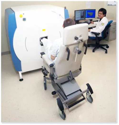
Scanco XtremeCT II (newest generation scanner)
- 3D In Vivo Human or Animal Imaging
- Knee Imaging capability
- Nominal Isotropic Resolution of 30 microns
Hologic Horizon A DXA
- Forearm, hip, spine imaging and osteoporosis classification
- Full body scans for body composition quantification
- TBS for vertebral quality evaluation
Imaging Capabilities
- Bone Morphometry and Skeletal Phenotyping
- Cancer and Vascular Imaging
- Biomaterials
- Osteoporosis Imaging
- Rheumatoid Arthritis Imaging
- Large-Scale Ex-Vivo Samples
- Multi-Modal Imaging (MRI)
Data Analysis and Management
The Bone Quality Research Lab is available to provide start-to-finish image analysis
and data management. Reach out to Faculty Director Galateia Kazakia, PhD to discuss
collaboration set up and fee structure.
- Morphometry
- Volumetric Quantification
- Segmentation
- Co-Registration
- Quantitative Parametric Maps
- 3D Visualization
- Custom Analysis Software Development
Quantitative Musculoskeletal Imaging Contact
Technical Director
Andrew Burghardt
Ph: (415) 514-9658
andrew.burghardt@ucsf.edu
Operations & Scheduling
Bo Fan
bo.fan@ucsf.edu