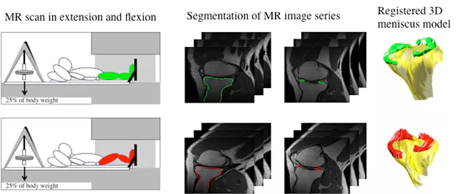
Osteoarthritis (OA) onset and progression is thought to be related to altered mechanical loading during daily activities. A fibrocartilagenous structure within the knee called the meniscus serves to distribute load across the joint to protect the underlying articular cartilage and bone. The meniscus is known to move as the knee flexes and extends, however abnormal meniscal movement during tasks such as walking and other activities of daily living may play an important role in the onset and progression of knee OA. Advanced quantitative magnetic resonance imaging (MRI) techniques may be used to evaluate meniscal motion, cartilage compositional changes, joint contact mechanics, and radiological grading of disease severity.
Therefore, the purpose of this project is to perform both cross-sectional (OA vs. controls) and longitudinal (3 times over two years) studies of meniscal motion in relationship to cartilage biochemistry. One hundred subjects with and without knee OA have been enrolled to complete kinematic-MRI and relaxation time mapping for cartilage and meniscus composition. Characterizing meniscal kinematics in participants with knee OA will improve out understanding of the mechanisms behind the incidence and progression of knee OA. This knowledge may be directly translated to the clinical setting since altered meniscal movement and morphology may be treated through surgical intervention, and targeting treatment specifically to those individuals that have the greatest risk for cartilage degeneration.
This research is supported by the National Institute of Arthritis and Musculoskeletal and Skin Diseases of the National Institutes of Health under Award Number 1R01AR062370 (PI: Souza) and 1F32AR064129 (PI: MacLeod). The content is solely the responsibility of the authors and does not necessarily represent the official views of the National Institutes of Health.