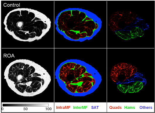
It is well known that obesity has a strong association with the risk of development and worsening of knee osteoarthritis. Weakness of the thigh muscles, especially the Quadriceps is a ubiquitous finding in people with knee OA. Loss of muscle tissue only partly explains the loss of strength in people with OA and fatty infiltration of thigh skeletal muscle is known to affect muscle strength and mobility in the elderly. However, little attention has been paid to the changes in fatty- infiltration of the thigh muscles in people with knee osteoarthritis. Using cutting-edge chemical shift-based water/fat separation MRI methods, including Dixon techniques and the iterative decomposition of water and fat with echo asymmetry and least-squares estimation (IDEAL), we are investigating the changes in thigh muscle size and adiposity in people with knee osteoarthritis. Furthermore, we are also investigating the association of thigh muscle size and adiposity with cartilage degeneration, muscle performance, and patient symptoms, function, and biomechanics. The project is currently funded by two grants from NIH-NIAMS (PI: Dr. Richard Souza, PI: Dr. Sharmila Majumdar).