Effects of reduced weight-bearing on skeletal geometry, micro-structure, strength
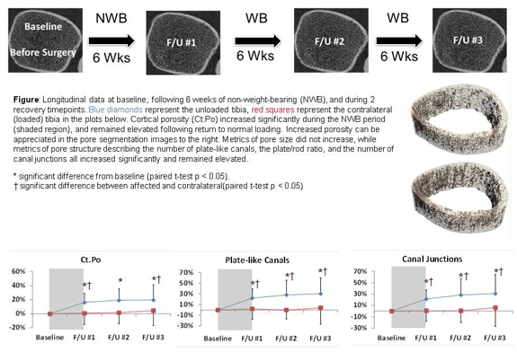
Longitudinal HR pQCT data acquired at baseline before surgery, after six weeks of non weight bearing, and during recovery at three follow up time points spaced six weeks apart. Blue diamonds represent the affected limb and red squares represent the contralateral limb. Non weight bearing resulted in increased cortical porosity that persisted during recovery. Segmented images show the non weight bearing region highlighted, with elevated porosity visible relative to normal loading. Quantitative metrics demonstrate significant increases in pore structure measures, including the number of plate like canals, the plate to rod ratio, and the number of canal junctions, which remained elevated over time. Statistical significance is indicated relative to baseline and between affected and contralateral limbs.
Significant bone is a common consequence of immobilization or restricted weight-bearing. Previous studies investigating bone loss in this context have focused on changes in bone mineral density (BMD). However, since cortical and trabecular microstructure contribute significantly to bone strength, we quantify and evaluate changes in microstructural and biomechanical parameters at the distal tibia during and after a period of non-weight-bearing. A long-term clinical focus of this research is to facilitate the development of targeted countermeasures to prevent bone loss.