Micro CT images comparing wild type and disease model bone, showing differences in cortical and trabecular structure, cross sectional views, and a graph of bone mineral density distribution highlighting altered mineralization.
Figure: Evaluation of bone quality in double transgenic (DT) compared to wildtype (WT) mice. Top:Full bone renderings (inset) and detailed sections from mid-diaphysis of femurs from 3-week old wildtype (left) and double transgenic (right) mice. Bottom: Mid-diaphyseal cross-section of a 9-week old double transgenic mouse (right) and SRμCT data (left) show a decrease in mineralized bone tissue and a disordered mineralization pattern.

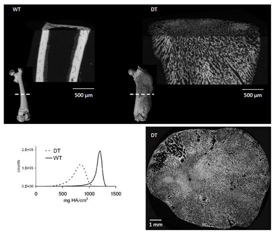
It is known that activation of the Gs G-protein coupled receptor (GPCR) pathway in osteoblasts can lead to significant increases in trabecular bone formation. However, the effects of constitutive Gs signaling on bone tissue quality are not known. Using Fourier transform infrared (FTIR) spectroscopy and synchrotron radiation μCT (SRμCT), we evaluate the bone composition of double transgenic mice that exhibit osteoblast-specific constitutive Gs signaling activity. Our findings demonstrate that Gs activity in osteoblasts leads to the deposition of immature bone tissue with reduced mineralization.