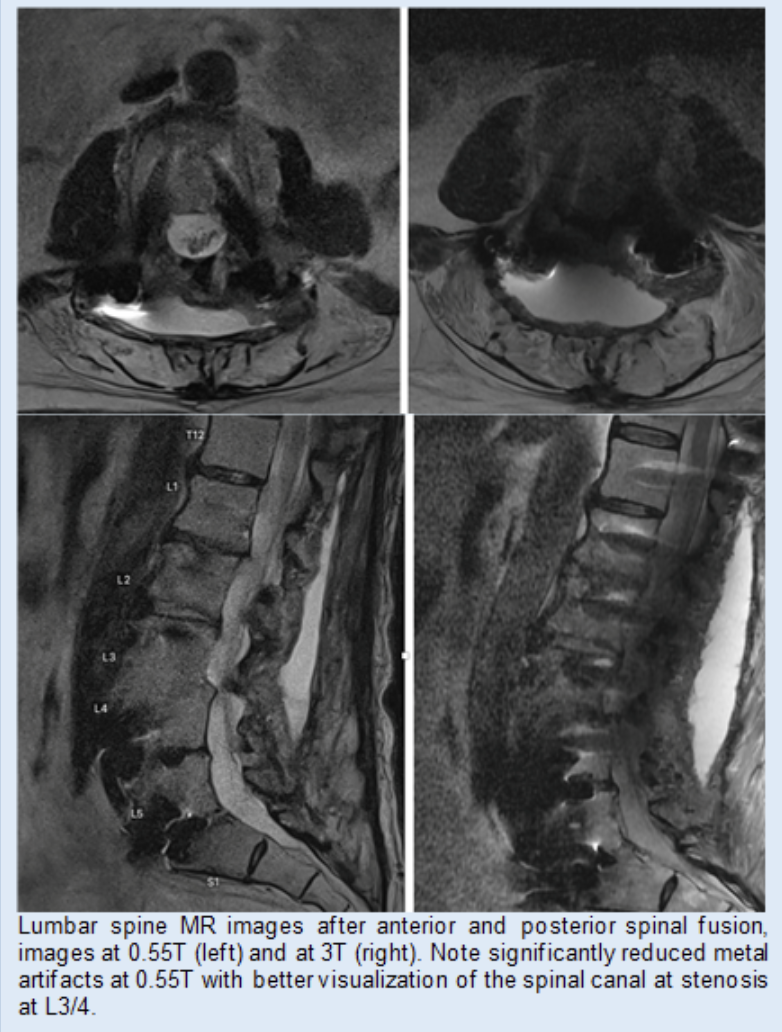
Using a novel 0.55T system we studied patients with metal implants at the spine (spinal fusion with metal hardware) and hip (hip arthroplasties) and found that imaging at 0.55T had superior image quality and showed abnormalities better than imaging at 1.5 or 3T. We also performed a biophantom study using metallic implants (steel and titanium screws) in pig knees which demonstrated substantial reduction of artifact size resulting in superior depiction of anatomical structures at 0.55 T MRI.
Read more about this project:
Improved metal suppression using new generation low-field MRI: a biophantom feasibility study. Luitjens J, Ziegeler K, Yoon D, Gassert F, Bhattacharjee R, Manatrakul R, Ngarmsrikam C, Becker A, Yang Y, Joseph GB, Su P, Itriago-Leon P, Majumdar S, Link TM. Skeletal Radiol. 2025 May;54(5):1093-1099. doi: 10.1007/s00256-024-04809-x. Epub 2024 Oct 4.