VA Advanced Imaging Research Center
The VA Advanced Imaging Research Center (VAARC) mission is to mirror the research structure of the Radiology Department in an effort to clearly define responsibilities in a democratic leadership framework and to promote teamwork as we strengthen and broaden our unique VA research program.
The VA Advanced Imaging Research Center (VAARC) performs clinical translational research on medical imaging with a focus on neurological and deployment-related illnesses and cardiovascular and musculoskeletal health of veterans. We aim to develop advanced diagnostic services to improve the health of veterans. An internationally recognized imaging research program, the VAARC consists of 35 scientists with an annual budget of more than $6 million from the National Institute of Health, Veterans Affairs, Department of Defense, non-profit foundations, and industry partners.
Research
The VAARC is a comprehensive research center that includes:
- Participant Recruitment
- Clinical and Cognitive Assessments
- Imaging Protocol Development
- Data Acquisition, Storage, Management, Processing, and Analysis
Staff diversity and expertise is our strength. Our research group includes clinicians, physicists, and computer scientists who focus on developing new techniques to improving our ability to acquire and analyze data. Our informatics team facilitates the project development and management, in partnership with out administrative personnel.
We live in an era where diseases are rarely conceptualized in a single system model, and fostering scientific collaboration among clinicians and specialty researchers is extremely important for the clinical translational research leadership and innovation and will ultimately to better serve our veterans.
Research Pillars
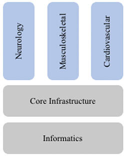 Focus on Three Clinical Pillars: We focus on Neurological, Musculoskeletal, and Cardiovascular biomedical imaging research where we can effectively impact the care of veterans, with infrastructure and informatics to support these clinical pillars.
- Core Infrastructure and Informatics Support: Advanced Imaging and Artificial Intelligence (AI) are critical technical strengths we offer under the CBI2 umbrella. We share these capabilities across our three clinical research pillars, as the technical tools have commonalities and synergies across medical areas.
Grants
The major goals of this Alzheimer’s Disease Neuroimaging Initiative (ADNI) are to:
- Develop improved methods, which will lead to uniform standards for acquiring longitudinal multisite MRI and PET data on patients with Alzheimer’s disease (AD), mild cognitive impairment (MCI), and controls.
- Acquire a generally accessible data repository, which describes longitudinal changes in brain structure and metabolism. In parallel, acquire clinical, cognitive and biomarker data for validation of imaging surrogates.
- Determine those methods, which provide maximum power to determine treatment effects in trials involving these patient populations.
The Dementia Research Group, London will perform image analysis of MR scans a) for the initial preparation phase of the study and support ongoing assurance of MR acquisition quality and stability and b) to provide atrophy rate measurements of all 0,6 and 12 month scans. Consistency of MR acquisition is essential for meaningful analyses of change. To this end each MR site will need to demonstrate that, in addition to performing scans of sufficient quality, they are also able to perform sufficiently reproducible acquisitions throughout the study. Each site will be assessed with registration of serial imaging of real subjects at the preparation/qualification phase and then again prior to every time point. Control, AD and MCI subjects will be rescanned at each site and the scan-rescan pairs will be accurately registered (positionally matched). Short interval scan pairs will be used to detect MR machine noise (same day scans), machine drift and operator consistency (1-2 week intervals). All registered scans will be analyzed to detect changes in acquisition parameters, image contrast and homogeneity or voxel dimensions and brain volume differences. Normative data will be used to define acceptable levels of variance and rates of change. In conjunction with other members of the consortium working on MR quality assurance, Dr Fox will work closely with the sites to correct problems that occur in terms of variation in acquisition. Registration-based measures of rate of brain atrophy and ventricular change (using the boundary shift integral) will be derived from all 200 AD and 400 MCI subjects and 200 controls for 0,6 and 12 month time point structural MR scans. Rates of change will be available for comparison with other MR and PET analyses and with the clinical measures. All pre-processing steps and brain regions derived as part of image processing will be made fully accessible.
VAARC Research Tools and Shared Resources
People
Directors


VAARC Steering Committee

In Memoriam

Contact Us
VA Advanced Imaging Research Center
Stephanie Rossi
Imaging Core Supervisor
San Francisco Veterans Affairs Medical Center
4150 Clement Street
San Francisco, CA 94121
Ph: (415) 221-4810 x26387
stephanie.rossi@ucsf.edu